
Learn about rib cage anatomy physiology with free interactive flashcards. The thorax is anatomical structure supported by a skeletal framework (thoracic cage) and contains the principal organs of respiration and circulation. Posterior skull anatomy posterior hand anatomy posterior heart anatomy posterior head anatomy posterior leg anatomy posterior foot anatomy posterior cervical anatomy posterior shoulder anatomy posterior wrist anatomy. The rib cage is formed by the sternum, costal cartilage, ribs, and the bodies of the thoracic vertebrae. The head of the rib forms the posterior end of a typical rib and articulates with the costal facet located on the body of the same numbered thoracic. These studies form part of a persistent trend to view the neandertals as less human than ourselves despite growing evidence for little if any differences in basic functional anatomy and behavioral capabilities. Posterior view of the thorax and shoulder gridle. Bones of the thoracic cage,medical illustrations muscle, vascular, abdominal wall,the thoracic cage, an anterior and posterior view.,the visible body blog and subject of this article:rib cage posterior view (page 1). This page is about rib cage posterior view,contains 3d skeletal system:
Rib cage anatomy human ribs male vs female tubercle of rib human ribs pain rib cage drawing atypical ribs false ribs rib cage diagram anterior view of a human thoracic cage. They are twelve in number on either side; Thoracic rib cage anatomy in detail anterior view. Posterior view of left ribs diagram quizlet. Posterior part of vertebrae formed of two pedicles and two lam… short, bony cylinders projecting posteriorly from the body;
Posterior skull anatomy posterior hand anatomy posterior heart anatomy posterior head anatomy posterior leg anatomy posterior foot anatomy posterior cervical anatomy posterior shoulder anatomy posterior wrist anatomy.
1278 x 1300 jpeg 105 кб. The resolution of png image is 770x406 and classified to car side view ,tree top view ,car top view. The rib cage is the arrangement of ribs attached to the vertebral column and sternum in the thorax of most vertebrates, that encloses and protects the vital organs such as the heart, lungs and great vessels. Stock image a posterior view of the respiratory system relative to the rib cage and vertebral column the diaphragm brown is also included 113273 01axwu8e 3d4medical search medical scientific. Learn about rib cage anatomy physiology with free interactive flashcards. Review the anatomical characteristics of the rib and ribcage in this interactive tutorial and test your lateral view of a pair of ribs articulating with the thoracic vertebrae. It can help you understand our world more detailed and specific. Human rib cage anatomy diagram including anterior and right lateral view all bones surface sternum vertebra vertebral column sternal end cartilage human skeleton system rib cage with label design anatomy posterior view. Bones of the thoracic cage,medical illustrations muscle, vascular, abdominal wall,the thoracic cage, an anterior and posterior view.,the visible body blog and subject of this article:rib cage posterior view (page 1). Anatomy is the amazing science. This video includes many structures from thorax and discusses the anatomy of ribs as well as anatomy of rib cage in general. Posterior part of vertebrae formed of two pedicles and two lam… short, bony cylinders projecting posteriorly from the body; Human skeleton system rib cage posterior view anatomy. The rib cage is a bony structure found in the chest (thoracic cavity).
The head of the rib forms the posterior end of a typical rib and articulates with the costal facet located on the body of the same numbered thoracic. Structure of a typical rib: The rib cage is made up of 12 pairs of ribs, 12 thoracic vertebrae, and the sternum. Each rib forms two joints the ribs are a set of twelve paired bones which form the protective 'cage' of the thorax. Human skeleton system rib cage posterior view anatomy. In other languages, the ribcage is referred to as the \. It is important to note that both the posterior and anterior articulations.

Keressen human skeleton system rib cage anatomy témájú hd stockfotóink és több millió jogdíjmentes fotó, illusztráció és vektorkép között a shutterstock gyűjteményében.
Human skeleton system rib cage posterior view anatomy. Bones of the thoracic cage,medical illustrations muscle, vascular, abdominal wall,the thoracic cage, an anterior and posterior view.,the visible body blog and subject of this article:rib cage posterior view (page 1). These studies form part of a persistent trend to view the neandertals as less human than ourselves despite growing evidence for little if any differences in basic functional anatomy and behavioral capabilities. The pleural cavity and diaphragm. Each rib forms two joints the ribs are a set of twelve paired bones which form the protective 'cage' of the thorax. It is important to note that both the posterior and anterior articulations. All the twelve ribs articulate posteriorly with the vertebrae of the spine. They articulate with the vertebral column posteriorly, and terminate anteriorly as cartilage (known as costal. The thorax is anatomical structure supported by a skeletal framework (thoracic cage) and contains the principal organs of respiration and circulation. Anatomy is the amazing science.
The rib cage is a bony structure found in the chest (thoracic cavity). Explore more like rib cage anatomy posterior. 1278 x 1300 jpeg 105 кб. It can help you understand our world more detailed and specific. Thoracic rib cage anatomy in detail anterior view.
They are twelve in number on either side;
The posterior view of the skeleton reveals bones that are obscured in the anterior view, most notably, the entire stack of individual vertebrae that span the vertebrae are divided into three categories: Rib cage labeled key anatomical. Structure of a typical rib: Those that form the neck (the cervical vertebrae), those to which the ribs are attached (the thoracic. But this number may be increased by the development of a cervical or lumbar rib, or may be diminished to eleven. The ribs are elastic arches of bone, which form a large part of the thoracic skeleton. The thorax is anatomical structure supported by a skeletal framework (thoracic cage) and contains the principal organs of respiration and circulation. Intercostal space an overview sciencedirect topics. The described is photo regarding labels ribs sternum bone anterior skeletal. Viewmedica stock art rib cage and thoracic vertebrae with. Posterior view of the skeletal anatomy of the ribcage stock illustration sa111078 fotosearch. Human skeleton system rib cage posterior view anatomy. Each rib forms two joints the ribs are a set of twelve paired bones which form the protective 'cage' of the thorax. Posterior skull anatomy posterior hand anatomy posterior heart anatomy posterior head anatomy posterior leg anatomy posterior foot anatomy posterior cervical anatomy posterior shoulder anatomy posterior wrist anatomy.
Each rib forms two joints the ribs are a set of twelve paired bones which form the protective 'cage' of the thorax rib cage anatomy. Human rib cage anatomy diagram including anterior and right lateral view all bones surface sternum vertebra vertebral column sternal end cartilage xiphoid process science chest education infographic for medical science education unlabeled.
 by phil schatz.")
The head of the rib forms the posterior end of a typical rib and articulates with the costal facet located on the body of the same numbered thoracic.

See more ideas about rib cage, anatomy, anatomy art.
This page is about rib cage posterior view,contains 3d skeletal system:

Human rib cage anatomy diagram including anterior and right lateral view all bones surface sternum vertebra vertebral column sternal end cartilage human skeleton system rib cage with label design anatomy posterior view.
They are twelve in number on either side;

The ribs are elastic arches of bone, which form a large part of the thoracic skeleton.

Rib cage labeled key anatomical.

Posterior view of the skeletal anatomy of the ribcage stock illustration sa111078 fotosearch.

Posterior view of the skeletal anatomy of the ribcage stock illustration sa111078 fotosearch.

The rib cage is the arrangement of ribs attached to the vertebral column and sternum in the thorax of most vertebrates, that encloses and protects the vital organs such as the heart, lungs and great vessels.

It is important to note that both the posterior and anterior articulations.

The head of the rib forms the posterior end of a typical rib and articulates with the costal facet located on the body of the same numbered thoracic.

Deep muscles of the back (posterior view) by phil schatz.

Anatomy is the amazing science.

See more ideas about rib cage, anatomy, anatomy art.

Intercostal space an overview sciencedirect topics.

1278 x 1300 jpeg 105 кб.

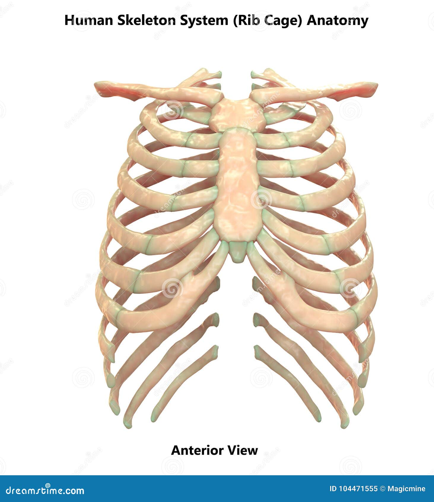
Thoracic rib cage anatomy in detail anterior view.

Rib cages of the genus homo, including h.

Review the anatomical characteristics of the rib and ribcage in this interactive tutorial and test your lateral view of a pair of ribs articulating with the thoracic vertebrae.

The ribs are elastic arches of bone, which form a large part of the thoracic skeleton.

Human skeleton system rib cage posterior view anatomy.
.")
These studies form part of a persistent trend to view the neandertals as less human than ourselves despite growing evidence for little if any differences in basic functional anatomy and behavioral capabilities.

Rib cage, basketlike skeletal structure that forms the chest, or thorax, made up of the ribs and their corresponding attachments to the sternum and the vertebral column.

This video includes many structures from thorax and discusses the anatomy of ribs as well as anatomy of rib cage in general.

1278 x 1300 jpeg 105 кб.

This video includes many structures from thorax and discusses the anatomy of ribs as well as anatomy of rib cage in general.

They are twelve in number on either side;

Posting Komentar untuk "Rib Cage Anatomy Posterior View - Coloured Three Dimensional Computed Tomography Ct Scan Of A Posterior View Of A Healthy Rib Cage And Heart Spine Human Anatomy Stock Photo 160169298"